Do you know RAMAN spectroscopy?part-3 / I will show you a used technique to be able to apply this phenomenon
Hello friends, lovers of science, today I return with a scientific publication always related to the area of materials science. While I have previously written about microscopy and many types of devices to characterize materials, today I will continue with the same line of publication with the third part of Raman spectroscopy, and in part I and II talk about the theory part and the main experimental bases to perform this type of microscopy, I did not show the proper technique to characterize a material, only in what it consists.
Now in this post if I want to write about a technique that is applied, talk about the basic mechanisms for this phenomenon related to Raman scattering to occur in this microscopy, there are many but it will start next:
credits of the image
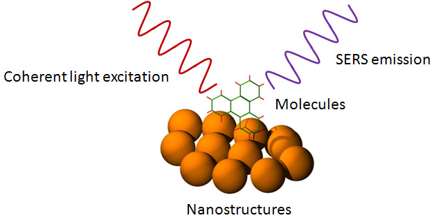
We can say that it is a spectral physical phenomenon by which light signals travel that are very weak, and that are related to the Raman scattering, what this dispersion does is to convert these weak signals into very powerful signals that can be detected in a very simple way.
The effect occurs when the plasmones are born on the surface of the material to be characterized by means of laser light to achieve a greater dispersion on the surface of the material.
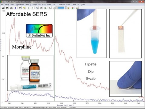
This technique uses a suspension in colloidal form of the metal, the Raman dispersion of the molecules that absorb on the surface of the metal causes it to be applied successfully, this technique allows to obtain analysis of very low concentrations of vibrations and spectra of the material, allows the detection of very small amounts of various substances.
As with traditional Raman spectroscopy, a monochromatic laser is used to produce the required dispersion. Before scattered light is analyzed, the most intense signal due to Rayleigh scattering is filtered to prevent overwhelming the Raman signals.
This technique uses two very important mechanisms for its operation, these depend on the nature of the molecules of the material and the experimental conditions and is given by the interaction between the molecule and the substrate [1].
Electromagnetic mechanisms (EM):
By striking a photon on the surface plasmons already located in the material, these palsmones are polarized from the small particle, through the resonance of the localized surface plasmon, resulting in it being the base of the intensification mechanism [2].

Scheme of the excitation of the plasmons located on the surface in a spherical particle by means of electromagnetic radiation.
credits of the image
The intensity of the Raman scattering signal occurs since the molecule does not affect only the electromagnetic radiation of the emission source, therefore also of the rough surface of the material to be characterized.
Because the intensity only depends on the magnetic field that falls on (I α E2), the signal occurs that is much higher than the signal that has the spectrum of the material, is produced by a considerable increase in the number of photons that affect the the characterized material. Therefore, if we have a greater number of photons, the probability that dispersion phenomena will occur increases, as well as absorption, and brings an increase in the effective section [2].
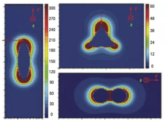
Intensifying fields around silver nanoparticles with an ellipse shape [3].
The image shows a considerable increase of the electromagnetic field, this field is heterogeneous, because it is located and concentrated in parts according to the geometry that the particles present, these points are known as hot spots, commonly found in spherical particles, if the particles are not spherical are located in areas of maximum curvature on the nanorrugosa surfaces [2].
Chemical or load transfer (CT) mechanism:
In this case when the molecule passes to the surface of the material, through a mechanism called chemisorption, a polarization occurs in the upper part of the adsorbate, thus creating states new electronics and a different symmetry. This helps a better electronic transfer between the metal and the orbitals of the formed material. After a certain time of relaxation this excited electronic state emits a lower frequency, granting a difference between the incidence and emission of the frequency of the arrival level and finally forming a resonance process [1].
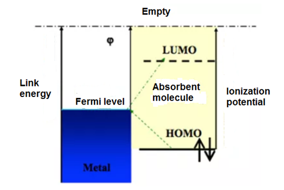
Load transfer processes between the metal and the material formed in the metal-adsorbate [1].
credits of the image
The most commonly used substrates are:
- Electrodes whose surface is rough by the application of various oxidation-reduction cycles.
- Metal islet films.
- Metal colloids.
- Lithographed nanostructures on a surface.
credits of the image

credits of the image
Through this technique we can observe the spectra obtained from different materials, more than any chemical substances, this technique may differ a bit from the characteristic Raman spectra, since the mechanism of signal intensification is mostly related to the CT.
Thus, the relative intensity of the spectrum bands can vary, due to the resonant effect of the plasmon and the complex formed; move to different wavelengths, due to the chemical changes that the formation of said complex produces on the molecule; widening of the bands, due to the appearance of new molecular states; and the appearance of new lines, because the strong gradients of the field give rise to different selection rules for this technique [4,5].
The other major difference, logically, is that the intensity of the Raman lines is greater than that found in conventional Raman spectra.
References:
[1]. Elena del Puerto Nevado. Detección y caracterización de quinacridonas de altas prestaciones mediante espectroscopías moleculares (Raman y fluorescencia) intensificadas por nanopartículas mecánicas. [Tesis doctoral]. Valladolid: Universidad de Valladolid; 2012.
[2]. http://eprints.ucm.es/17911/1/T34150.pdf
[3]. Haynes CL, McFarland AD, Van Duyne RP. Surface-Enhanced Raman spectroscopy. Analytical Chemistry. 2005; 77 (17): 338 A-346 A.
[4]. Kneipp K, Kneipp H, Itzkan I, Dasari RR, Feld MS. Surfaced-enhanced Raman scattering and biophysics. Journal of Physics: Condensed Matter. 2002; 14: R597- R624.
Other sources:
https://web.archive.org/web/20071202054629/https://www.andor.com/chemistry/?app=64
http://www.prucommercialre.com/que-es-la-surface-enhanced-raman-scattering/
http://www.horiba.com/fileadmin/uploads/Scientific/Documents/Raman/RA08.pdf
Thank you very much to the @steemstem team for the support received and the acceptance to your community.