THE ANATOMY OF HUMAN TOOTH:THE UNDER-RATED PART OF HUMAN BODY
Hello friends! Hope you all are having a great time.
Today we will be discussing something about one of the most under-rated parts of human bodyi.e.our pearly whites or the teeth. Hope you will find it useful.

source-pixaby
IMPORTANCE OF TEETH:DO WE NEED TEETH ONLY TO EAT?
Here are the functions of teeth. Apart from the well known funtion of mastication,it has several other importances.
- Help in mechanically breaking down the food. Humans are omnivorous and feed on both plants and animals so our teeth are designed that way. The incisors help in cutting or biting food items like carrot,radish,bananas and other food items into small pieces. Likewise,canines help in ripping and tearing the food especially meat and the premolars and molars help in chewing and grinding the food. Thus we can enjoy all varieties of food.
- Helps in speech by forming words by controlling airflow through the mouth.Some sounds are made when tongue strikes against the teeth.for example-the sound "th" is made when tongue strikes against the upper row of teeth.
- Gives shaape and beauty to the face.
- Helps in generating facial expressions.
- Owing to their highly resistant nature and intact structure even many years after the death of a person, they have a special place for identification in forensic medicine in a branch called forensic odontology.
- Can be used for age estimation of a person and also the unique bite marks can be used for identification.
- In some other animals, they can be used for defense and attack.
General discussion:
We all know that life is not impossible but very difficult if we don't have teeth because of its functions as mentioned above.Human beings are diphyodonts i.e. they replace their teeth only once in their lifetime.
The initial set of teeth that we have are the temporary/milk/baby/deciduous teeth which are 20 in number and are later repalced by the 32 permanent teeth.
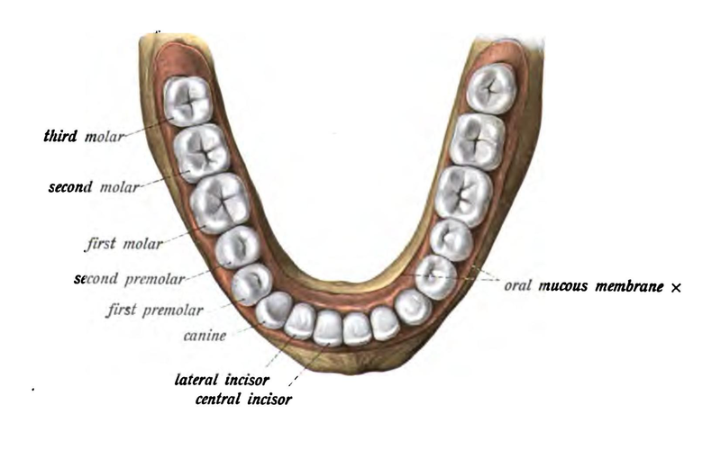
source by Dr. Johannes Sobotta
The following consist of the 20 temporary teeth.
- Incisors-8
- Canines-4
- Molars-8
And the following make up the 32 permanent teeth. - Insicors-8
- Canines-4
- Premolars-8 and
- Moalrs-12
The first teeth to erupt is the lower central incisor at around 6 months of age and the last teeth to erupt is the third molar ,also known as the "wisdom teeth" that may appear between 17-25 years of age but in some people they may don't appear at all. There is a period of mixed dentition when both primary and permanent teeth teeth are present together in the oral cavity. It is seen between 6-12 years of age from the time when the first permanent teeth appears to the time when the last temporary teeth sheds.

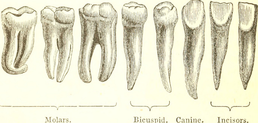
source by Henry Gray
Teeth may have one or more roots. Usually incisors,canines and most premolars have only one root with the exception of maxillary pre-molar that usually has 2 roots along with mandibular molars. Maxillary molars on the other hand have three roots.
sourceby Carpenter,William Benjamin
Now let's discuss the parts of human tooth.
At first, to make the discussion easier let's divide tooth into:
1.Crown-The part of the tooth seen in the mouth after eruption and is covered by enamel.
2.Cementoenamel junction,dental cervix or the neck-The part of the tooth where the cementum covering the root and the enamel meet each other i.e.the junction between root and crown.
3.Root-Part below the neck that is attached to the alveolar bone by the periodontal ligament.
Now let's discuss in a bit detail.
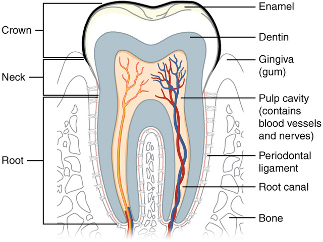
Toothby,Open Stax College,CC by 3
a.Enamel:It is the hardest and most mineralised substance of the body consisting of 96% minerals, chiefly hydroxyapatite Ca10(PO4)6(OH)2, called as crystalline calcium phosphate. It provides strength and brittleness to the enamel. Remaining 4% of the enamel is composed of water and organic materials. Enamel is semitranslucent so the color of enamel is highly affected by the color of underlying dentin and any restorative materials if used. Normally it may be yellowish to greyish white in colour.
The rate of wear of enamel, also known as attrition, under natural conditions is around 8 micrometers/year.
Medical imporatnce
- The enamel is highly protective against wear and tear of the teeth.
- Because it is insensitive(lacks nerve supply), small cracks and pits in enamel are painless.
- Dissolution of enamel by the poor dental hygiene and lots of carbohydrates leads to bacterial build up, formation
of acid and dental caries which are extremely painful.
b.Dentin:Dentin is the mineralised connective tissue with organic matrix of collagenous protein consisting of 70%inorganic material, 20%oragnic material and 10% water by weight.It is covered by enamel on the crown and the cementum on the root and surrounds the entire pulp. The process of formation of dentin known as dentinogenesis is initiated by the odontoblasts of the dental pulp.
Dentin supports the enamel. The differntiating features between enamel and dentin are that the latter is sensitive and formed throughout the life and is softer and decays more easily. Small microscopic channels called dentinal tubules are present throughout the dentin that radiate from pulp cavity to the exterior enamel or cementum border.
Medical importance
- Exposure of the dentin due to tooth decay, tooth-brush abrasion or gum recession can cause sensitivity in the tooth due to the microscopic nerve fibres present in the dentin.
c.Cementum:It is specialised bone like substance covering the root of the tooth consisting of 45% hydroxyapatite, 33% oraganic material, chiefly collagen and 22% water. It is secreted by cementoblasts within the root of the tooth and is softer than both enamel and dentin. The chief function is to help in the attachment of periodontal ligament to the tooth.
Medical importance
- Recession of gums at old age can lead to the abrasion of cementum due to its low mineral content and thinness leading to exposure of the deep dentin and problems like extrinsic staining and dentinal hypersensitivity.
- Cementicles are small spherical or ovoid calcified masses attached to or embedded within the cementum layer or attached to periodontal ligament.
d.Dental pulp:It is the unmineralised oral tissue consiting of soft connective tissue and neurovascular elements that occupies the central pulp cavity of each tooth. It has soft gelatinous consistency and the major component both by weight or volume is water. The pulp cavity extends downwards through the root of the tooth and opens outside at the apical foramen. This forms communication between pulp and surrounding tissue that helps in the spread of inflammation from pulp to the surrounding. Cells like odontoblasts that form dentin along with cells like fibroblasts, macrophages, lymphocytes are present in the pulp. Also the matrix consisting of collagen and the ground substance rich in glyprotein, proteoglycans and water is present.
The major function is formation of dentin by odontoblasts along with nutrition, sensation and defence.
Blood vessels entering the pulp through the apical foramen branch extensively and peripherally to form a dense capillary network just beneath or sometimes extending into the odontoblast layer. These capillaries have pores that represent the high metabolic activity of the odontoblasts.
Small venules drain the capillary bed and leave as vein through the apical foramen.
Many nerves enter the tooth via the apical foramen of the premolar and molar tooth with single nerve entering the foramen of other teeth. The nerves have primarily two modalities.

Nerve supply of tooth-1.enamel 2.dentinal tubule 3.dentin 4.odontoblastic process 5.predentin 6.odontoblast 7.capillaries 8.fibroblasts 9.nerves 10.artery/vein 11.cell-rich zone 12.cell poor zone 13.pulp chamber,by Ian Frust,CCby3
a.Autonomic nerve fibres:
Only sympathetic nerve fibres are present in the pulp. These nerve fibres extend from neurons whose cell bodies are present at the superior cervical ganglion at the base of skull. These are unmyelinated fibres and travel along with blood vessels supplying their smooth muscles and thus help in the regulation of blood flow inside the blood vessels.
b.Afferent(Sensory) fibres:
These arise from the maxillary and mandibular divisions of the fifth cranial nerve i.e.the trigeminal nerve. These are predominantly myelinated fibres and terminate in the central pulp. From this region, some will send out small individual fibers that will form the subodontoblastic plexus of Raschkow just under the odontoblast layer. From this plexus, fibres extend in an unmyelinated form towards the odontoblasts. The fibres terminate as free nerve endings near the odontoblasts, may extend between them or may reach even upto dentinal tubules. They transmit pain stimuli from heat, cold and pressure. It is mainly located in the roof and lateral wall of the coronal pulp(pulp present in the crown) and less developed in the root canals.
Medical importance
- Inflammation of pulp is called pulpitis which is very painful . Due to the closed environment around the pulp, the pressure increases with inflammation which is extremely painful and finally leads to the compression of nerves and necrosis of pulp.
- As the pulp becomes exposed due to caries or any other reasons, extreme pain occurs due to the nerves present.
SUPPORTING STRUCTURES:
Periodontium is the supporting structure of tooth helping the tooth to attach to surrounding tissues. It consists of cementum, periodontal ligaments, alveolar bone and gingiva.
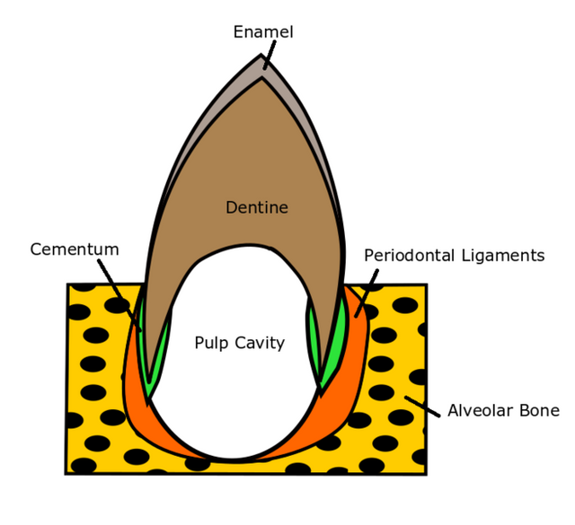
supporting structures,by Dinohk,CC by4
a.Periodontal ligament-
It is the specialised connective tissue that connects cementum of root to the alveolar bone. Its functions are attachment of tooth to bone,formation and resorption of tooth during tooth movement, sensation and eruption.
Cells include osteoblasts, osteoclasts, macrophages, cementoblasts etc.It is a neurovascular structure so during chewing or biting, when pressure is applied to the tooth,the tooth moves slightly in the socket putting a tension on the periodontal ligament from where the information is sent via the nerves to the CNS for interpretation.
Clinical significance
- It plays an important role in healing after trauma or surgery.
- It can be used for giving anaesthesia for local pain control in child and adolescents.
b.Alveoalr bone-
It is the thickened ridge of bone that forms the tooth socket on bones that hold the teeth.In humans, the teeth bearing bones are the maxilla and mandible. The alveolar bone undergoes modification throughout the life by the activity of osteoblasts and osteoclasts.
Clinical significance
- Alveolar bone is lost due to periodontal diseases.
- Bone is stimulated by chewing and speech.If teeth are lost,the stimulation is also lost and bone is resorped.
c.Gingiva:
It is the mucosal tissue that overlays the mandible and maxilla inside the mouth. Healthy gingiva is pink in color with smooth curved contour and firm texture.
Clinical significance
- Gingival recession can lead to exposed root surface which is very sensitive.
- Improper oral hygiene can lead to gingivitis.
- Disturbed structure of gingiva is aesthetically unacceptable.
So teeth are an important part of our body and lack of proper oral hygiene can lead to dental caries which are extremely painful and cost a lot of money. In USA, dental caries are at third place just after heart diseases and cancer in terms of annual medical expenditure on hospital visits.
So keep your pearly whites healthy.I will be discussing dental caries in my next blog and it was the anatomical base for that article.
AT LAST, I WOULD LIKE TO THANK @scienceangel from the bottom of my heart who cleared all my doubts and helped me a lot to edit the article .
Also I would like to thank @chloroform for his valuable feedback.
References:
http://www.uky.edu/~brmacp/oralhist/module4/lecture/oh4lectm.htm
https://www.mouthhealthy.org/en/az-topics/t/tooth
https://en.wikipedia.org/wiki/Human_tooth
https://en.wikipedia.org/wiki/Pulp_(tooth)
https://www.webmd.com/oral-health/picture-of-the-teeth#1
https://www.britannica.com/science/tooth-anatomy
If you liked it,you can upvote,resteem and comment.
Any queries regarding the post are heartly welcome.
Hi @ameet77804. There are a few issues with your article:
Really, is it the most underrated part? I have heard about the navel being as one of the most underrated body parts. I'm not sure about teeth.
There are quite a number of typos. You might want to recheck before clicking the post button in the future.
If you have any problem regarding STEM-related articles, you can join steemSTEM Discord Channel and we will be glad to assist you.
@chloroform
I really tried a lot not to land in the copyright issues again as in my previous article.
@mobbs had tried telling me about the copyright issues in the previous article.SO this time,I used the pictures with my best knowledge not to commit any mistake again.
I had read the link given by you before posting this article today and I even tried looking at the trending posts in steemit how they use the images.
I am so sorry that I unknowingly breached the rules.I accept that I had some spelling mistakes that I have corrected and as far as considering teeth as underrated is concerned,I just meant to say that people don't pay as much imporatnce to teeth as they should because the disease of teeth are highly prevalent all over the world.
I still have no clue how my citing of the images is incorrect.Please help me if you can.
Thank you so much for your kind feedback.
I assumed this issue was resolved in the Discord channel.
Yes sir and a I have completely edited my article as well.
Resteemed your article. This article was resteemed because you are part of the New Steemians project. You can learn more about it here: https://steemit.com/introduceyourself/@gaman/new-steemians-project-launch