The Evolution of Medical Imaging
The medical field is quite a field that has experienced a vast growing development over the years. The imaging part of the medical world has undergone a series of evolutionary improvements to satisfy the standards of efficiency of clinical diagnosis and treatments of various diseases. Science and technology has really helped the medical world to attain its height of reputation and effectiveness. What would have been said about medicine if the magnificent contributions of scientific and technological innovations were absent. However, when did the evolution begin and how has it grown to attain its present state of development.
Original Image source
Edited by @okunlolayk
The science of medical imaging is a very important arm of the medical field and it serves as the first line of action in diagnosing any abnormality in the body system. Medical imaging is a clinical procedure that involves the visualization and capturing of the interior tissues and organs without having to open the body. Through medical imaging, we are able to peer into the interior of the human body to diagnose, treat and monitor various ailments. The imaging technology has saved physicians a lot of time and stress, as they could now see the interior of the body without surgically opening through. This imaging gives the visual representation of the normal anatomy and physiology of the body and makes it easy to identify abnormalities. However, let’s trip down the timeline of the evolution of medical imaging.
The revolution was birthed at the beginning of the 20th century when Professor Wilhelm Conrad Roentgen accidentally discovered X-ray. He was working on Cathode ray tube when he noticed that the invisible rays could penetrate solids of different densities, differently. At this moment he doesn’t know what these rays are, so he called them X-rays. This discovery gives rise to the oldest medical imaging technique known as X-ray. Roentgen made the first X-ray by achieving the image of his wife’s left hand. During this period, capturing with X-rays involves the projection of an amount of radiation through the body to focus on a piece of sensitive film inside a cassette. The patient is exposed to this radiation for about 11minutes to get an image on the X-ray film. This technique depends on the differential absorption of the X-ray radiation by the various tissues of the body on its way to the sensitive film. The film being sensitive to the exposure of radiation creates an image outline depending on the amount of radiation reaching it. When the x-ray is projected, the radiation is absorbed by anything that stands it way. However, the level of absorption depends on the density of the material. Bones and teeth are the most absorbent tissue, body fluids are intermediate while air is least. Therefore, each point on the radiograph (film) has a density (blackness) which is determined by the amount of x-rays reaching it. The film is deeply blackened outside the area obstructed by the body and appears clear due to absorption in regions where the penetration was occluded. This film, however, undergoes a wet developer process in a dark room to aid visualization.
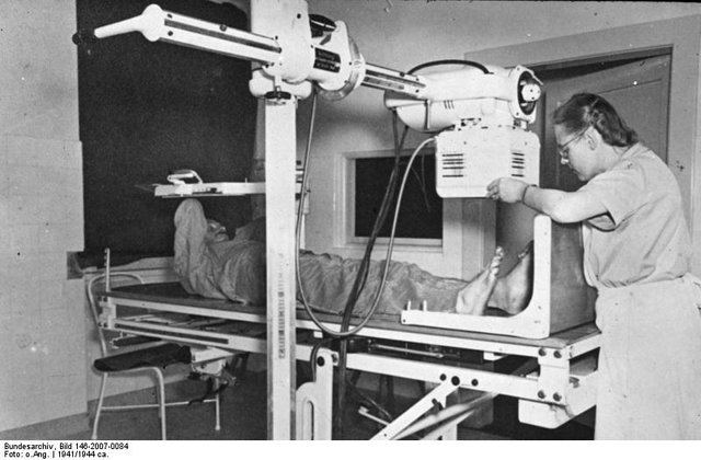
An X-ray examination of the Leg, 1941
Image Source:wikimedia
Attribution: Bundesarchiv, Bild 146-2007-0084 / CC-BY-SA 3.0
Sooner to this period the Fluoroscopy technique was invented. With this technique, we could not only image bony structures but now capture hollow organs by introducing a dense contrast medium into them. Fluoroscopy involves the use of a radio-opaque substance (such as barium) as a material that creates a dense contrast as it travels down hollow organs like the Gastrointestinal tract (GIT). This radio –opaque substance, when introduced into hollow organs, it absorbs the penetrating radiation and makes the organ visible as the substance travels the tract. This procedure is used in the diagnosis of cancers, ulcers, diverticulitis and appendicitis which are disorders of the GIT.
With the knowledge of X-ray, a number of new techniques were birthed. Mammography is also an imaging technique that was built on the knowledge of projective x-rays. It is a technique that takes high-resolution images of the breast to diagnose breast cancer. Angiography is also a product of radiology, which is a technique used in taking the image of blood vessels. Just like fluoroscopy, Angiography involves the injection of a radio-dense contrast substance into the artery of interest in order to get an image of it. Later soon, in 1950, the Selinger technique from Karolinska institute was adopted. In this technique, a flexible guide wire is inserted through a puncture needle and a plastic catheter is passed over the guided wire and directed towards the organ of interest. This imaging technique has made possible to diagnose vascular malformation, vessel narrowing or blockage.
During the late 40s, Positron and artificial radiation were discovered as well as recognition of short-lived radio-nuclides in tracing biochemical and physiological pathways. In the 1950s, nuclear medicine was introduced in the practice of diagnostic imaging. In this form of imaging technique, X-ray tubes are not used for the radioactive projection but rather
a radioactive substance that emits gamma rays as they decay. This radioactive substance is introduced into the body of study and the radiations are detected by special gamma cameras. However, the nuclear scan is used in the detection of the spread of cancer and tumors. As the science of nuclear medicine begins to boom, Institutions like universities, hospitals, and health societies begin to make research to build on the founding basis of nuclear medicine and employ its knowledge in biomedical studies. This led to the invention of the PET.
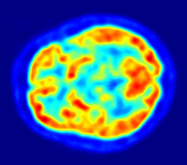
Positron Emitting Tomography (PET) is an imaging procedure that involves the detection of signals produced by the annihilation of a positron and an electron yielding two photons of 511KeV travelling in opposite directions. Unlike the early nuclear procedures that emit gamma rays, PET makes use of isotopes that decay to emit positrons (a negatively charged electron). A deeper attention was paid to the use of short-lived isotopes because the majority of life-sustaining metabolic processes occurs within a very short period of time. However, cyclotrons are designed with flexibility to produce isotopes with half-life short enough to trace metabolic pathways. At first, PET makes use of the detection of annihilation radiation over a single photon but within a short period, there was advancement from the single photon technique to a double photon technique. The double photon technique makes use of 511 KeV photons moving in opposite direction and in coincidence with reference to the source of action thereby creating a more defined image. The development of positron cameras also improves the sensitivity and resolution of the PET.
In the early 1970s, computer technology was introduced to the field of medical imaging and this led to the advent of Computer Tomography (CT scanning). CT was a momentous advancement in imaging technology as it allows multiple tomographic images to be obtained digitally. Before the advent of CT, X-ray images were taken on plane films which could only show the position and displacement of tissues (especially bones). However, CT allows the X-ray tube to be rotated at different angles around the area of interest and the detectors pick up the measure of penetration as the radiation passes through the body tissues.
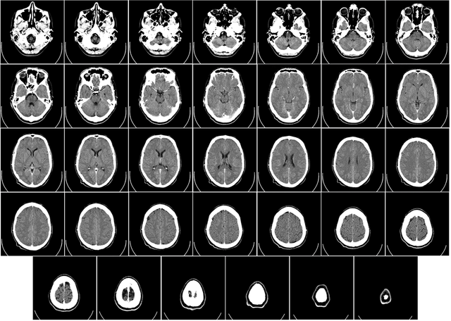
A Computer Tomographic scan of the Human Brain, from base of skull to tpp
Image Source:wikipedia liscence: CC0
The CT machine was invented by Sir Godfrey Hounsfield who made a theory that the image of an object can be seen if the X-ray is taken from different angles of the object through a machine that would appear as slices which could be put together to form an image. Sir Godfrey ideology forms the concept of Axial Tomography.
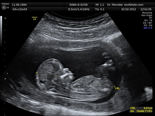
Ultrasonography also appeared in the 1970s. This is a unique imaging technique because it does not involve the use of projective radiation or radio-dense contrast material like the earlier techniques.
Ultrasound is an imaging technique characterized by the passing of a beam of high-frequency sound waves with low wavelength through the body to hit the organ/ tissues of interest. Based on the composition of this target tissue the waves reflect or bounce back to the detector to create tomographic images of the characteristics of the tissue. This is a non-invasive and non-radioactive technique in which the body is not subjected to the harmful effects of any ionizing radiations. This characteristic makes this technique ideal for the imaging of delicate tissues, and fetus during pregnancy.
Magnetic Resonance Imaging (MRI) or Nuclear Magnetic Resonance (NMR) is also an imaging technique that evolved during the 1970s. Just like ultrasound, it does not require the use of ionizing radiation but function by creating a strong magnetic field to produce an image of spatial resolution. An MRI scanner consists of a large and powerful superconducting magnet with a 100cm bore and creating a uniform magnetic field. The magnetic field produced is about 1.5 telsa which is 30,000 times stronger than the earth’s magnetic strength. However, due to this magnetic field surrounding the object, certain atomic nuclei absorbs and emits radiofrequency energy that can be interpreted by the machine to produce a well detailed image of the object. MRI is a versatile imaging technique that is not only restricted to the medical field but also useful in chemistry to take images of both organic and inorganic structures (as in NMR spectroscopy).
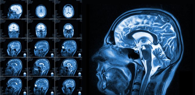
Magnetic Resonance Image of the Brain
Image source
In the clinical test, the radio signals are emitted by hydrogen proton. This is because there is abundant hydrogen in body fluid composition. This proton produces two phaces of signals: first signal(T1) which is detected when the when the protons are excited by the environmental magnetc field and the second signal (T2) which is detected when the protons return back to their normal state when the magnetism is off. However the detector receives both signals (T1 & T2) and by processing their voltage difference, it produces the image of the object. Also because the protons in different tissues generate signals differently, the machine is able to create an image of these tissues with distinctions. The MRI technique is used generate an image of both anatomical structure and physiological processes.
Today, X-ray images are now acquired digitally without any film. Fluoroscopy is still in use but it has advanced considerably with the use of modern image intensifier and sometimes being replaced by Computer Tomography. The old X-ray tomography has now metamorphosize into an advance Computer Axial Tomography scanning. PET is now being used with CT forming PET-CT to acquire better and more detailed metabolic information and a high spatial resolution. The PET machine designs also shows improvement in geometry; from Planar, to Polygonal to circular geometry. In big medical centres, the large volume of medical images are now being managed with PACS. Picture Archiving and Communications System (PACS) is a medical imaging management technology that makes it easier to store, access and manage images in the radiology department.
Conclusion
There has been a great evolutionary advancement since the onset of the medical imaging science. Over the last century to this moment, the medical imaging field has shown a fast trend of advancement which has really benefited the armamentarium of the medical world; there has been a great reduction of in hazards associated to the various imaging tests, production of 3D images, high resolution images, minimal time taken for the procedure and efficiency of the overall procedure. However, these improvements are not just credited to the efforts of the medical field only but also the contributions of other fields of science and technology as well as efforts of dedicated scientists.
References
- http://www.imaginis.com/faq/history-of-medical-diagnosis-and-diagnostic-imaging
- https://en.wikipedia.org/wiki/Medical_imaging
- http://www.umich.edu/~ners580/ners-bioe_481/lectures/pdfs/2008-09-procAmerPhilSoc_Bradley-MedicalImagingHistory.pdf
- https://en.wikipedia.org/wiki/Magnetic_resonance_imaging
Referential Source links for this article are included for further reading and all Illustrative Images are attributed to their various sources. Thank you.
Best Regards!!

Hello! I find your post valuable for the wafrica community! Thanks for the great post! We encourage and support quality contents and projects from the West African region.
Do you have a suggestion, concern or want to appear as a guest author on WAfrica, join our discord server and discuss with a member of our curation team.
Don't forget to join us every Sunday by 20:30GMT for our Sunday WAFRO party on our discord channel. Thank you.
thanks a lot for your support
This post has been voted on by the SteemSTEM curation team and voting trail in collaboration with @curie.
If you appreciate the work we are doing then consider voting both projects for witness by selecting stem.witness and curie!
For additional information please join us on the SteemSTEM discord and to get to know the rest of the community!
Congratulations @okunlolayk! You have completed the following achievement on the Steem blockchain and have been rewarded with new badge(s) :
Click here to view your Board of Honor
If you no longer want to receive notifications, reply to this comment with the word
STOP