Reptile and Amphibian X-Rays
One of the coolest features of our facility is our Animal Health and Surgery Suite. Under the supervision of our resident veterinary technician, the surgery suite is where we perform the countless routine procedures, rehabilitation and medicating that keeps our animals in top shape. Thanks to generous contributions from vets, doctors, hospitals and other medical facilities, we even have our own ultrasound, sonogram and x-ray machines that allow us to keep an eye on our patients from the inside. These are just a few of the reptile and amphibian x-rays we have taken on site!
Note: Generally, x-rays and other animal medical records are not open to the public and are not allowed on social media. However, these particular x-rays have been approved for public use and are featured our childrens' "Be a Veterinarian" play area, allowing kids a chance to experience a hands-on approach to wildlife medicine.
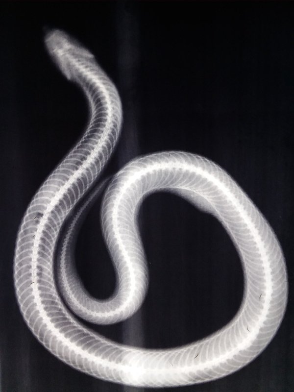
Eastern Hognose Snake:
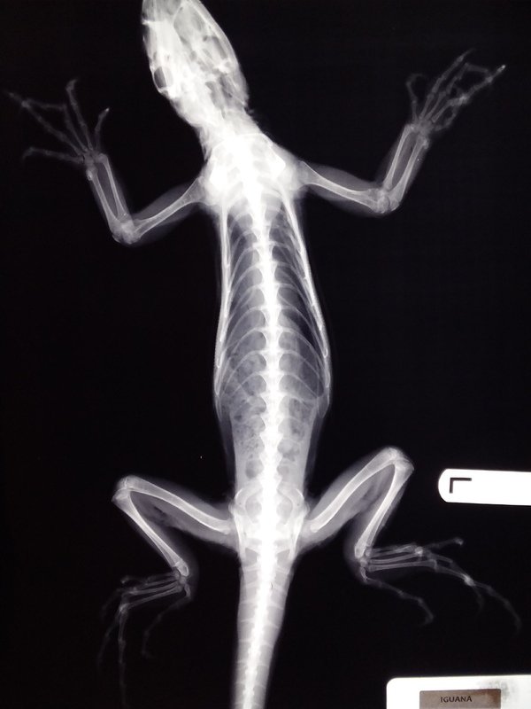
Green Iguana:
This iguana came to us a few years ago after it was surrendered by an owner who didn't know how to properly care for it. It suffered from a pretty severe case of metabolic bone disease (which you can see most clearly in the fingers). Since the x-ray was taken, the iguana has been put on a specialized diet to help mitigate the damage and now has a happy new home with a more knowledgeable owner.
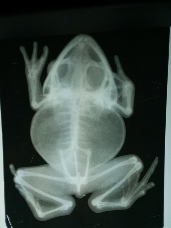
American Bullfrog:
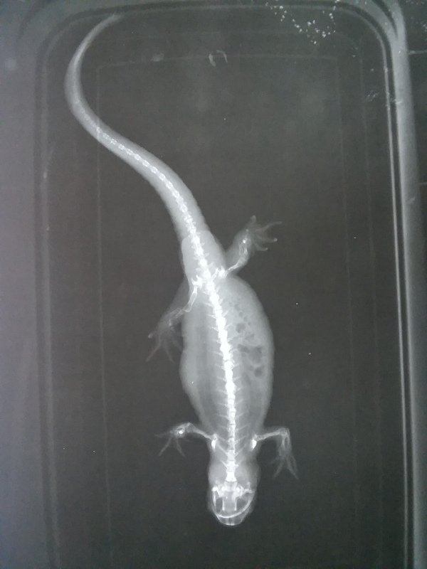
Eastern Tiger Salamander:
Bearded Dragon:
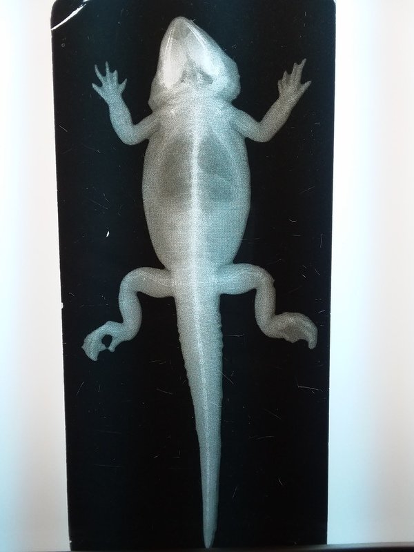
Northern Copperhead:
You may notice the lines on either side of the snake. When working with venomous snakes (hots) we put them in a plastic tube to avoid being bitten while we work.
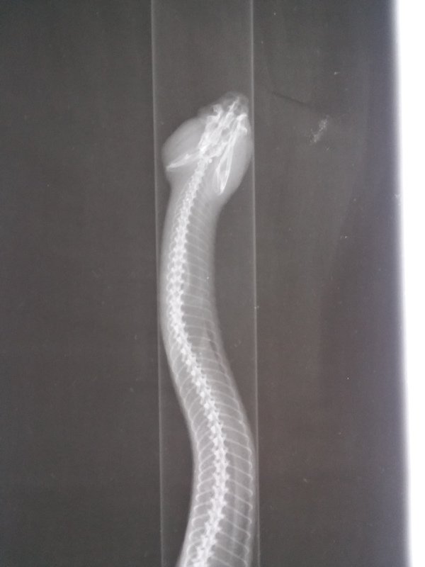
American Alligator:
X-raying an alligator is definitely tricky. Besides trying to get the animal to cooperate, they have osteoderms that make them difficult to x-ray. Osteoderms are bones embedded in the skin, and trying to scan through these to get an accurate picture of the skeleton is difficult to say the least.
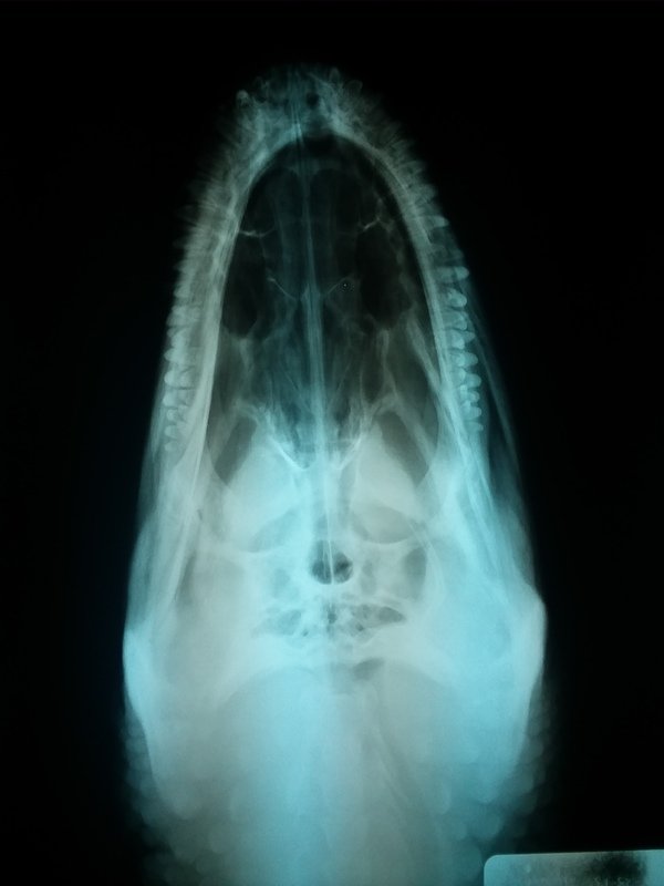
Leopard Tortoise:
The tortoise below is named "Tiki" and is not a part of our collection. Tiki was one of 7 tortoises that were rescued from a pet store fire a few years ago (unfortunately they were the only survivors). They were admitted to our facility for treatment; they all had inhaled quite a bit of smoke and were having respiratory difficulties. But Tiki's x-ray revealed something more surprising than the damage to her lungs: she was carrying eggs! Since the treatment, Tiki and the other tortoises have found new homes.
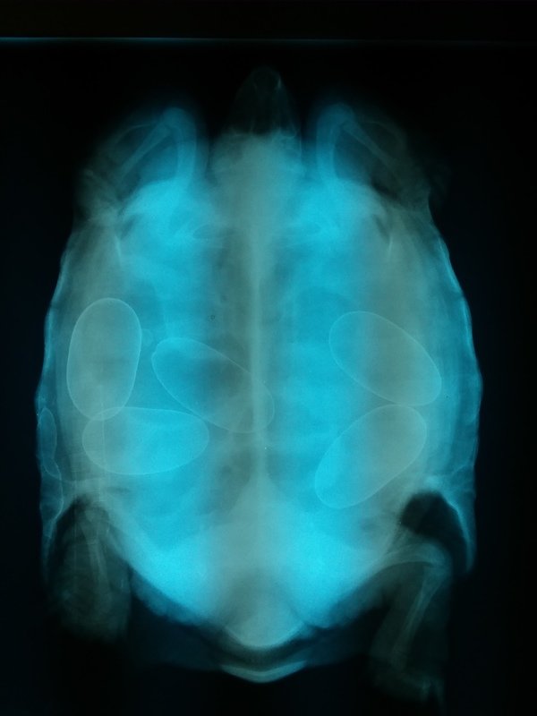
These are pretty interesting images. What can one learn from the Xray?
Quite a bit if you know what to look for. Obviously bone conditions like the iguana above and pregnancy like the tortoise, but we can also check for growths and tumors that may suggest cancers or other diseases. We can even see fluid levels within the lungs that can alert us to issues like pneumonia. If we suspect an animal has consumed something it shouldn't have, we can check the stomach for foreign bodies without having to resort immediately to surgery.
WOW
Awesome, love the eggs in the tortoise, she looks rather full.
Very very interesting. Never seen reptiles and amphibians through an X ray.