The Aesthetics of Anatomical Illustration
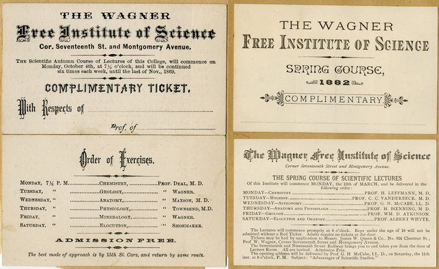
Human anatomy & physiology was a staple of the Wagner’s educational program during its first thirty years. It was taught almost every semester between 1855 and 1885 by over 15 different professors. One of the more frequently employed professors was Dr. Ralph M. Townsend, who taught at the Wagner beginning in 1868 for almost a decade. Prof. Townsend was a graduate in the 1866 class at Jefferson Medical College where he later became Professor of Anatomy. He was also a Fellow at the College of Physicians of Philadelphia and wrote extensively on medical and surgical practices.

Course announcements for Fall 1869 and Spring 1882 semesters. From the Scrapbooks of the Wagner Free Institute of Science.
Human anatomy and physiology were the foundation of a medical education, but little of the population could attend these schools. The Wagner’s courses, taught by doctors, provided access to information about the human body that few had access to. And according to an 1868 entry made by William Wagner in the Annals of the Wagner Free Institute of Science,
Human Anatomy & Physiology on Wednesdays attracted larger audiences than any other subjects.

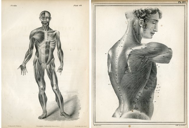
Plates depicting the muscular system.
- Left: Quain, Jones. A series of anatomical plates; with references and physiological comments, illustrating the structure of the different parts of the human body. Philadelphia : Carey and Hart, 1843. MAIN 611 17.
- Right: Cloquet, Jules. Atlas du Manuel d’Anatomie Descriptive. Paris : Béchet, 1825. MAIN 611 19.
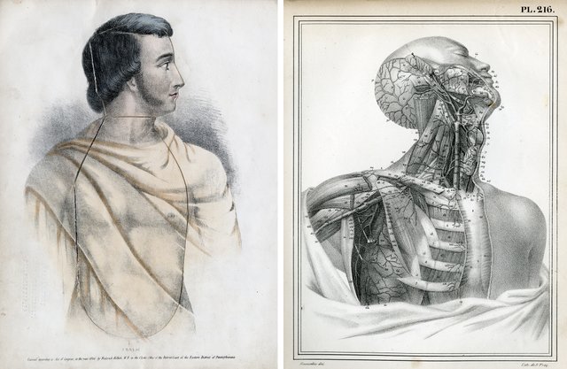
In medical school, these courses would have been taught using anatomical specimens and human cadavers. These were not available at the Wagner, so courses were illustrated by large, hanging teaching charts and books filled with detailed anatomical illustrations. These illustrations, drawn with such skill and detail, can also be appreciated for their artistic merit.

- Left: Hollick, Frederick. Outlines of anatomy & physiology : illustrated by a new dissected plate of the human organization. Philadelphia : T.B. Peterson, ©1846. MAIN 611 13.
- Right: Cloquet, Jules. Atlas du Manuel d’Anatomie Descriptive. Paris : Béchet, 1825. MAIN 611 19.
Temple University-Wagner Fellow, Natalia Vieyra, a PhD student in Art History at Tyler School of Art, Temple University, used the Wagner’s collection of 19th century anatomy and physiology texts as part of her ongoing thesis project Under the Skin: Anatomical Illustrations and the Aesthetics of Medical Knowledge. Her project delves into the interdisciplinary aspects of these books, considering the intersection of aesthetics, visual culture, science, and medicine.
Ms. Vieyra’s project
examines mid-19th century anatomical illustrations, investigating their unique function as both transmitters of scientific information and objects of aesthetic interest, as well as placing them in relation to period discourses surrounding questions of race, gender, and nationalism is 19th century America.
In the Wagner’s collection, Ms. Vieyra was especially interested in the Classical imagery used in some of the anatomical illustrations. In reference to the fold-out anatomical diagram in Hollick’s Outlines of Anatomy and Physiology, she states,
The figure’s muscular physique, lightly veiled by neoclassical drapery, signifies a conception of vigor and health that is rooted in a Classical past in keeping with the mid-century American identification with Classical Republican values.
Video of the fold-out anatomical diagram in Hollick’s Outlines of Anatomy and Physiology.
All books are from the collection of the Library of the Wagner Free Institute of Science.
Ms. Vieyra is continuing her research at other libraries and plans on publishing her conclusions in the next year.
100% of the SBD rewards from this post will support the continuation of a fellowship program at the Wagner Free Institute of Science.
