FAMILY LIFE EDUCATION & EMERGING HEALTH ISSUES. @dansanctus
A study of the human reproduction and development is a study of the mode of development of the generative organs. How human life begins is very complex and interesting. Beginning from a simple one cell organism know as a zygote, the individual grows through a process of cell division, unto a stage-by-stage process of maturation into adulthood.
The human cell result from the union of a sperm cell from the male and an ovum (egg) from the female, during sexual intercourse. This fused single cell carries. 23 pairs of chromosomes which carry the genes responsible for every development in the human being. These genes bear all the hereditary traits of the individual. These traits include the colour of the eye, skin, hair, blood type, height and weight of the individual.
THE REPRODUCTIVE ORGANS.
The anatomy of the reproductive organs means the study of the structure of the organs while the physiology deals with the functions of the organs. The reproductive organ of the male is the penis while the female is the vagina. These are the two noticeable sex organs used by human beings for reproduction.
The Male's Organs of Generation (The External Parts and Their Functions).

A. THE PENIS
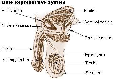
This is the most noticeable part of the male genitals. It is a boneless organ which is made up of spongy tissues. It is covered by the foreskin which a part of it is removed during circumcision. The skin has no hair. It is loosely arranged to allow for expansion in erection. It resemble a tube with a tip called the glans at the end of it. The penis is connected to the body through what is called the root. The raised ridge is called the corona (crown). The coronal ridge and the glans are the most sensitive region for sexual stimulation in the male. At the tip of the penis is an opening know as the urethra through which urine and semen pass. The penis serves important functions in reproduction, excretion of waste product by urination and in sexual pleasure. The penis is connected with the vas deferens and the prostrate gland from within.
The penis is between 6.4cm (2.5 inches) width and 10cm (4 inches) long when flaccid. When aroused or erected it is much longer (roughly between 15cm (6 inches) width and 33cm (13 inches) long.
B. THE SCROTUM (SCROTAL SAC)
The scrotum sac is the pouch of loose skin that hangs below the penis. It contains the testes. It is situated below the public bone and just beneath the penis. The sac protects the testes and controls the temperature necessary for sperm production and survival.
THE INTERNAL PARTS AND THEIR FUNCTIONS
A. THE TESTES.
This is called testicles. They are two in number, they are the reproductive glands in males, like the ovaries in the females, they produce sperm cell and sex hormones called testosterone. Both testes are about the same size but the left side usually hangs lower then the right side. The scrotum and the testes move up and close to the body when the weather gets too cold, and away from the body when there is rise in the temperature. This helps the sperm to survive in whichever way the temperature changes.
B. EPIDYMIS
The epidymis is a small network of coiled tubes behind the testes where the sperm mature. They are temporary store for immature sperm until they attain full maturation and are released upon ejaculation when they are passed from the epididymis into the vas deferens.
C. VAS-DEFERENS
These are the tiny tubes that carry the sperm cells from the epididymis into the seminal vesicles. Each vas deferens leads into the ejaculatory duct which opens into the urethra. Sperm cells are ejaculated out through the penis via the urethra.
D. SEMINAL VESICLES
These are small sac-like structure found around the urethra. They produce the fluid that forms part of the semen. The fluid produced helps to nourish the sperm cells. The sperm cells have little motility of their own while in the epididymis and the vas deferens until they mix with the seminal fluid.
E. THE PROSTRATE GLAND
The prostrate gland lies below the bladder. It is made up of both muscles and glandular tissues. It is fairly small at birth, enlarges at puberty and possibly shrinks in old age. It produce a milky and alkaline fluid called the prostatic fluid which forms part of the ejaculate. The alkalinity of the fluid helps to prevent the destruction of the sperm cells by the acidity of the female vagina. Thus, prolonging the life span of the sperm in the female reproductive system. The secreted fluid combines with the seminal fluid giving the semen a characteristic texture and colour. The prostrate gland can enlarge causing an interference with urination. In this case, the person requires a surgical or a medical therapy.
E. COWPER'S GLAND
The cowper's glands are found one on either side of the urethra. It produces a clear alkaline fluid at the tip of the penis especially during sexual arousal, just before ejaculation occurs. It is meant to neutralize the acidic urethra in the male and allow the safe passage of the sperm. Fluid secreted by this gland sometimes carries some stray sperm capable of making the woman become pregnant even when the man has not ejaculated.
G. THE URETHRA
The urethra runs from the neck of the bladder and opens to the exterior at the tip of the penis. It serves as a passage way for the urine and the sperm.