The art and science of dental radiographs (Part 1)

Like a doctor is handicapped without a stethoscope, a mechanic without a screwdriver, and a soccer player without their boots, a dentist is equally handicapped without a radiograph, commonly referred to as an X-ray. A radiograph is one of the most essential tools in dentistry, as it provides a clear and detailed view of what lies beneath the surface. Without it, a dentist cannot reach an accurate diagnosis, and without a proper diagnosis, making a treatment plan becomes impossible. Without a solid treatment plan, addressing and treating the patient's ailments effectively is almost impossible.
Radiographs play an important role in detecting hidden dental issues, such as cavities between teeth, infections, bone loss, impacted teeth, or other abnormalities that are not visible during a clinical examination. They serve as a window into the unseen, guiding dentists toward making informed decisions and delivering optimal care.
In this post, I will discuss the common findings and interpretations of dental radiographs. By the end, you will have a basic understanding of what to expect when looking at an X-ray. So, the next time you visit your dentist, you’ll have a clearer picture of what you’re looking at and how it relates to your oral health.
Case 1 |
|---|

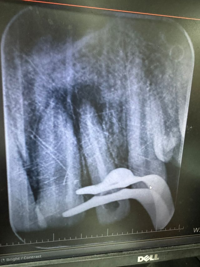
This radiograph is one of the clearest I have taken recently, and it highlights a tooth affected by decay. Let me explain it in simpler terms while also using some dental terminology for clarity.
In this image, you can see a tooth with visible decay on its top surface, known as the occlusal surface. The darker area visible in the radiograph represents the decayed portion, also called "radiolucency." This happens because decay, or caries, causes the tooth structure to break down, which appears as a dark shadow on an X-ray.
Inside every tooth, there’s a living part that consists of nerves and blood vessels. This central part is called the pulp. The pulp has two main sections: the coronal pulp, which is located in the crown (the visible part of the tooth), and the radicular pulp, which extends down into the roots. When decay progresses and gets very close to or reaches the pulp chamber, the tooth can become sensitive or even painful. At this stage, treatment is required to save the tooth.
If the decay is very close to the pulp but hasn’t entirely invaded it, dentists may perform a procedure called direct pulp capping. This involves placing a protective material over the pulp to seal it and prevent further infection. However, if the decay reaches the pulp chamber, the tooth becomes infected, and a root canal treatment (RCT) is necessary. In an RCT, the infected pulp is removed, and the space is cleaned, filled, and sealed to save the tooth from extraction.
Additionally, in this radiograph, you can observe an area of radiolucency around the tip of the tooth’s roots, called the periapical region. "Periapical" refers to the area surrounding the apex or tip of the root. This radiolucency is a sign that the infection has spread beyond the tooth into the bone around the root. This condition is often referred to as periapical pathology or a periapical abscess, and it typically occurs when the infection within the tooth has traveled down through the root canals into the surrounding tissues.
To summarize, this radiograph shows a tooth with advanced decay affecting its internal structures. While the decay has reached the pulp chamber and caused infection at the root tip, treatments like root canal therapy or pulp capping can help preserve the tooth and restore its function. This image serves as a great example of the importance of early detection and timely treatment to prevent complications.
Case 2 |
|---|

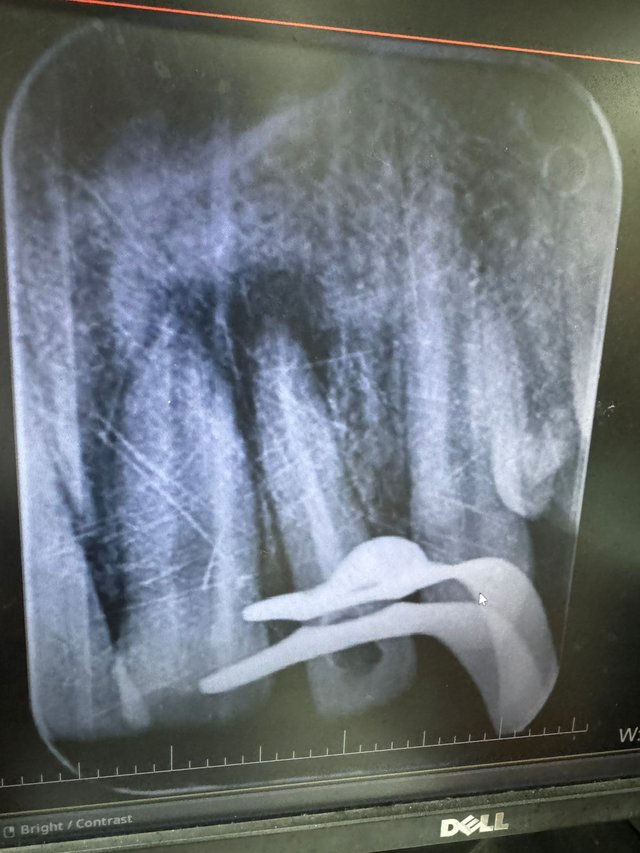
This radiograph belongs to a 17-year-old boy who experienced trauma during childhood, affecting his front teeth (anterior teeth). The image shows two adjacent teeth, with one of them filled with a white material called Gutta Percha. This material is used to seal the root canal system during a Root Canal Treatment (RCT). The adjacent tooth, however, appears empty, with no such filling.
If you observe closely, there’s a large radiolucent area (dark shadow) around the root tips (apices) of both teeth. Radiolucency in this region often indicates an infection or inflammation in the bone around the root apex, commonly referred to as a periapical lesion. This occurs when bacteria from a damaged or decayed tooth enter the surrounding tissues, causing an abscess or infection.
The most striking feature of the empty tooth is its open apex—an incomplete root formation with the tip of the root remaining open. This condition is typically caused by trauma or infection in younger patients while the tooth is still developing. Trauma can disrupt the normal growth and closure of the root, leaving it immature and open. This condition not only makes the tooth more fragile but also poses a challenge during treatment because it is difficult to seal the open apex effectively.
For this patient, the infection was severe, as evident by the large radiolucent area. To address this, I performed a Root Canal Treatment (RCT) on the affected tooth. This procedure involved cleaning out the infected pulp (the soft tissue inside the tooth containing nerves and blood vessels) and disinfecting the canal to eliminate bacteria. The next challenge was to close the open apex. For this, a material called Mineral Trioxide Aggregate (MTA) was used. MTA is a bioactive material that can seal an open apex effectively. It promotes the formation of hard tissue and provides a tight seal to prevent reinfection.
The radiograph shown here was taken mid-procedure, and you can see a wing-like structure in the image. This is the rubber dam clamp, which is used to isolate the working field during dental procedures. Isolation ensures that the tooth stays dry and free from saliva, which could compromise the treatment.
From a dental perspective, this case highlights the complexities of managing immature teeth with open apices, especially when combined with a significant periapical infection and if I were to explain in simpler terms, the patient had a severely infected tooth with an underdeveloped root tip due to childhood trauma. I treated the infection by cleaning the tooth and sealing the root using specialized materials, which allowed the tooth to heal and function normally over time. Follow-up radiographs revealed that the infection subsided significantly, and the bone around the root began to regenerate, indicating a successful outcome.
Case 3 |
|---|

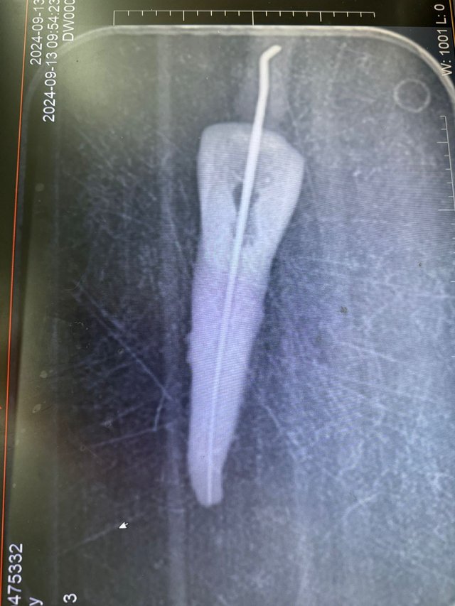
This is a radiograph of an extracted tooth, commonly used for teaching and practice purposes in dentistry. It serves as a demonstration tool, particularly for procedures like filing during root canal treatments. The tooth appears isolated in the image because it is no longer in the oral cavity but rather outside the mouth for educational purposes.
Filing is a critical step in performing a root canal treatment. It involves the use of specialized small instruments called files, which are typically made of nickel-titanium or stainless steel. The purpose of filing is to clean and shape the root canals by removing the infected or necrotic pulp tissue and debris. This prepares the canal for sealing and prevents further infection.
Dentists carefully use files to clean and shape the canal to its proper taper. One of the fundamental rules of filing is to respect the anatomy of the canal. Dentists must avoid perforating the canal walls or extending the instrument beyond the apical foramen (the tip of the root). A perforation or overextension could lead to treatment failure, persistent infection, or complications like pain or swelling.
This was it from the Art and Science of Dental Radiographs (Part1)
If you learned something new from this article or have any questions, do let me know in the comments section.
Regards,
Dr Huzaifa Naveed
There are so many things involved in having a healthy mouth. It's easy to see why people have always had issues keeping their teeth.
True. This comment made me think of maybe starting a series of 'why did this happen?' where I'll show a problem and discuss all the possibilities which led to that specific problem
Hi, @huzaifanaveed1,
Your post has been manually curated!